Our skin is the largest and one of the most important organs in our body. It not only serves to regulate our health, but also plays a decisive role in our well-being.
The tasks of the skin range from the first line of defense against bacteria and viruses to the regulation of fluid balance and body temperature.
This highly sensitive organ can serve as a mirror of health, which is why both skin diseases and other illnesses can have a negative effect on the appearance of the skin.
Find out everything you need to know about the structure and function of the skin and why it’s worth taking intensive care of your skin health.
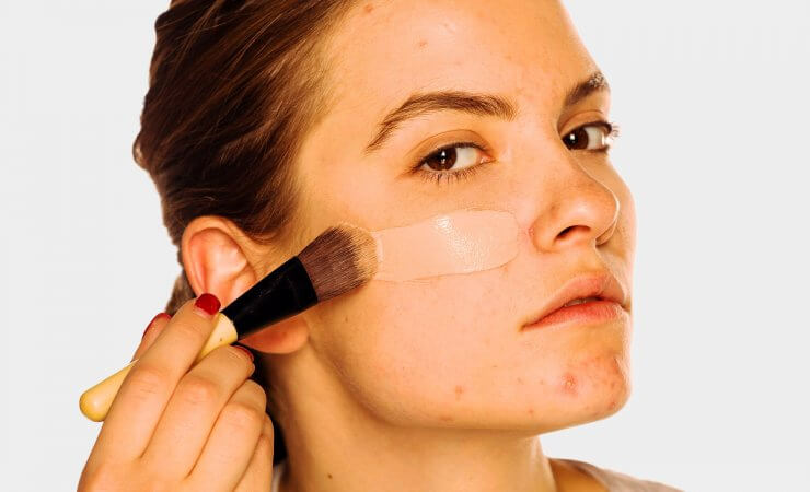
Many people around the world, especially teenagers (15 to 18 years) and young adults, suffer from the skin condition acne. Pimples, blackheads and redness are typical acne symptoms.
With the right treatment, however, they can be reduced to a minimum. In this article you will learn everything you need to know about acne, its causes and treatment.
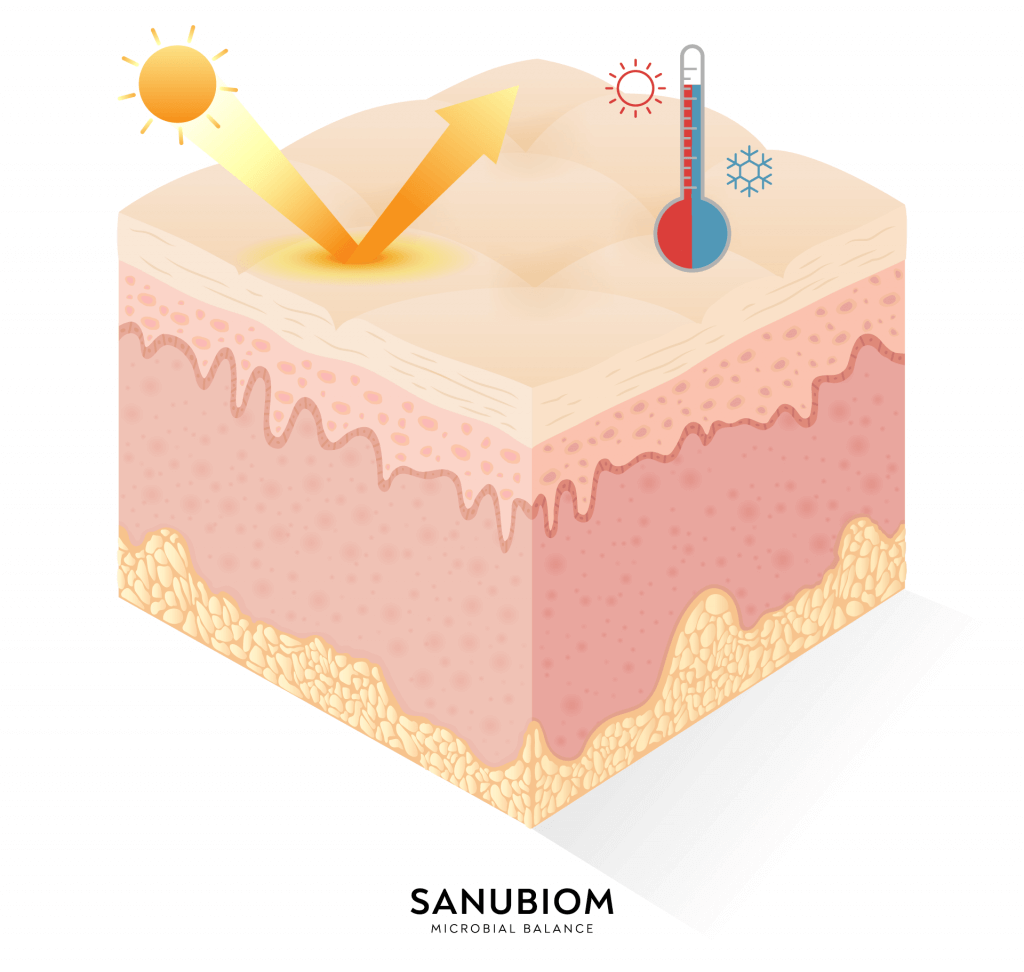
The skin is the largest organ in our body. With an area of one and a half to two square meters, it envelops our organism and protects it from external influences.
It is only one to two millimeters thick, but still weighs around three and a half to ten kilograms.
Depending on the amount of blood, the proportion of pigment-forming cells and the thickness of the top layer, each person has an individual coloration and composition of the skin.
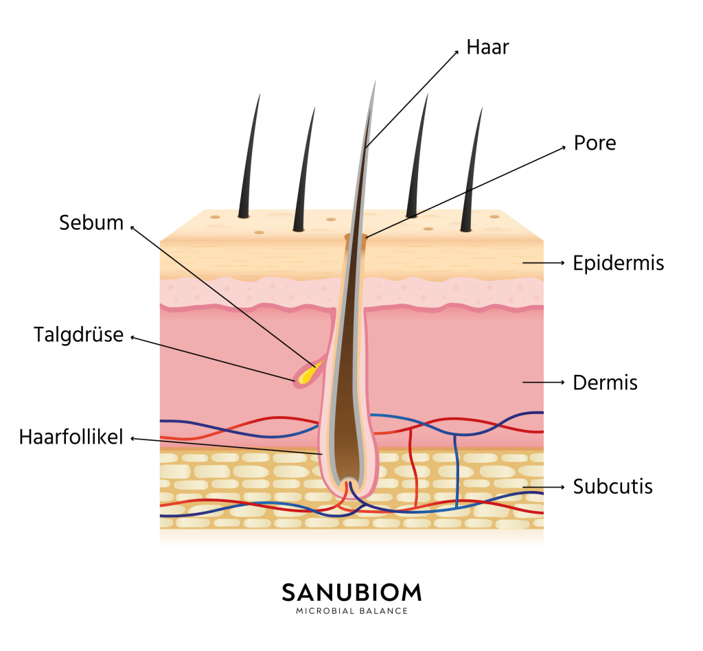
The structure of the skin consists of 3 layers. In addition to the outer epidermis, we also have a dermis and the deepest subcutis.
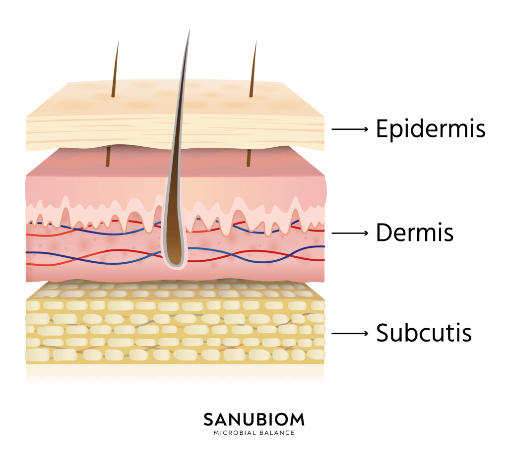
The epidermis is the uppermost of 3 layers of the skin. It consists largely of a horny layer, which is made up of special cells that keratinize on the surface and are then shed.
In this way, the epidermis is constantly renewed. The epidermis is also made up of individual layers. These include:
The dermis (also known as the corium) is the middle layer of the skin.
It lies between the epidermis and the subcutis and consists mainly of connective tissue fibers.
It is itself divided into two layers, which are not really separated from each other, but merge seamlessly:
In addition to sebaceous and sweat glands, the dermis also contains hair follicles, blood and lymph vessels, nerves and muscle cells.
The subcutis is the lowest of the three skin layers. It consists of closed connective tissue chambers that are filled with fat cells.
It is firmly connected to the overlying dermis by strong fibers of connective tissue and also has mechanical contacts with the underlying structures.
The uppermost section of the subcutis has sweat glands, the deeper parts of the hair follicles and receptors of the skin (so-called Vater-Pacini corpuscles).
Skin appendages are all structures that are formed from the epidermis or dermis and are located in the skin.
The skin appendages include
The physiology of the skin is vital for the survival of our organism. It protects against external influences such as pathogens, heat or UV radiation and enables sensory impressions to be perceived and transmitted to the brain.
Without the skin, no movement would be possible for us. It offers mechanical protection and should not be damaged, even if it is subjected to heavy pressure.
Overall, it can be said that the skin is most resilient where muscles lie underneath as a cushion. Where the skin runs directly over the bone, however, it is extremely sensitive.
As the top layer of the epidermis is consumed under stress, it is continuously renewed.
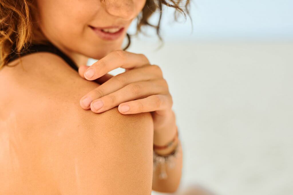
The top layer of skin is able to absorb UV rays. If the UV-B radiation lasts for a longer period of time, this horny layer thickens and a so-called light callus is formed, which prevents the dangerous UV radiation from damaging the deep unkeratinized skin layers.
Moreover, deeper penetrating rays are almost completely absorbed by the melanin pigment and converted into heat. The result is the much-loved “vacation tan“.
If someone becomes very tanned, this simply means that the skin has produced more melanin in order to better protect itself from UV radiation.
One of the most important properties of the skin is its protection against pathogens. If it is injured or weakened, pathogens can otherwise penetrate and trigger dangerous infections.
The focus here is on Langerhans cells. These cells are located in the stratum spinosum of the skin and resemble macrophages, i.e. specific immune cells. They are therefore an essential part of our natural defense system.
Scientists are therefore conducting numerous studies on the interaction of skin cells with immune cells to defend against pathogens.1
Every day we need our skin for sensory perception. In addition, there are the so-called sensory corpuscles of the skin, through which various sensory qualities are perceived.
The most important are free nerve endings, Merkel cells, Ruffini corpuscles and Vater-Pacini corpuscles.
In this way, mechanical and thermal stimuli, vibrations and pain, for example, can be perceived and transmitted to the brain as information.
Maintaining body temperature is particularly important. The blood vessels in the papillary layer in the dermis are responsible for this. These vessels expand at high temperatures.
As they are very close to the surface, the blood can cool down and the body temperature is lowered. Conversely, the blood vessels contract when it is cold and prevent the blood and the body from cooling down further.
Itchy skin, redness and sprouting pimples under the skin characterize the appearance of patients with a skin disease.
Every skin is different and depending on your skin type, individual habits or life circumstances, your skin can be susceptible to a wide range of skin conditions. However, the range of possible diseases is huge. We have summarized the 8 most common ones for you.
Acne is a very common skin disease, especially during puberty, which is caused by a predisposition. The sebum in the skin cannot drain properly through the pores because they are clogged or keratinized.
Patients subsequently suffer from blemished skin with reddish inflammations and pus-filled pimples. The conspicuous appearance of the skin can be an enormous burden, especially for adolescents.
Atopic dermatitis is particularly common in (young) children. The skin disease can occur all over the body, with the elbows and hollows of the knees being particularly affected.
Itchy skin that is red and flaky are typical symptoms that are caused by excessively dry skin.
The first signs of rosacea are visible blood vessels on the nose and cheeks. In addition, there are small pustules and papules which, in the worst case, can develop into a coarse connective tissue skin pattern.
Perioral dermatitis usually occurs in women in the form of reddened skin around the mouth and nose, leaving a symptom-free border directly around the lips.
In addition, small, painful pimples can appear that do not simply heal by themselves, but can remain for months.
Melanomas are primarily recognized by altered moles. These can change in diameter, color or elevation and thus be a sign of black skin cancer.
As the risk increases with age, you should have annual check-ups with a dermatologist from the age of 30 at the latest so that surgery can be performed as early as possible in the event of an emergency.
Most skin diseases are not contagious. However, athlete’s foot does pose a risk, which is why bathing shoes should always be worn, especially in public pools.
With athlete’s foot, the affected skin between the toes softens and appears swollen. In addition, the affected areas often itch or hurt. Nail fungus, on the other hand, often triggers a change in the shape, color or thickness of the toenail.
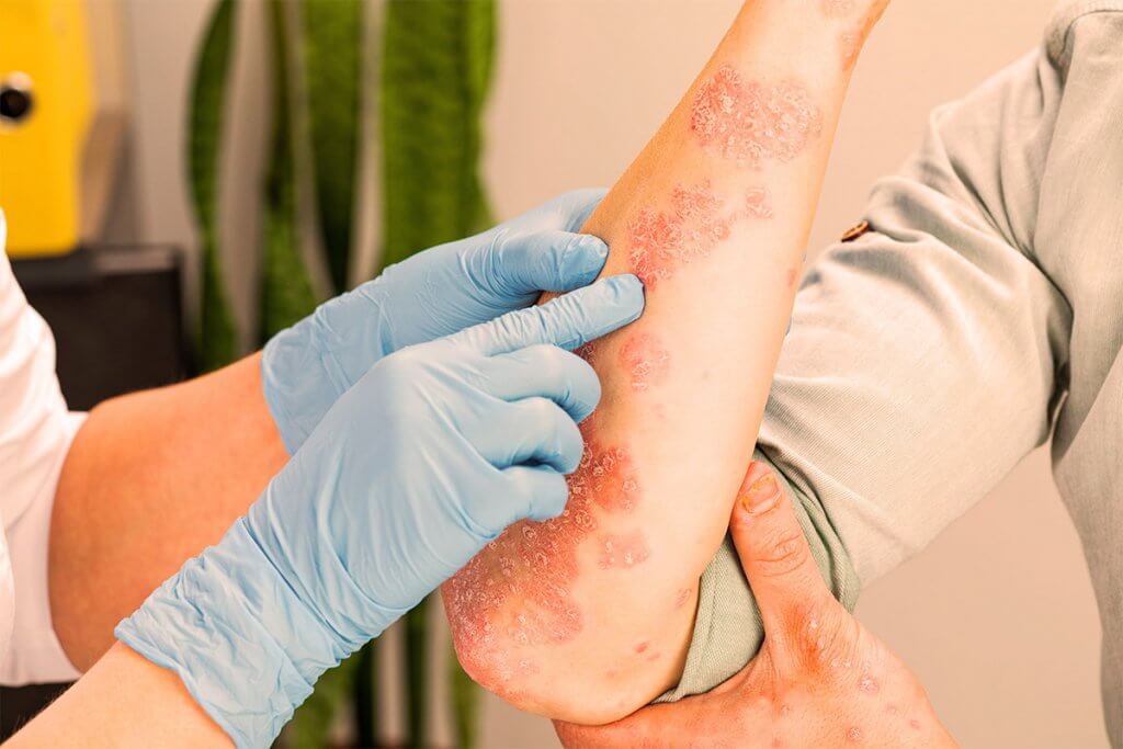
In the case of psoriasis, the skin becomes red in individual areas such as the elbows, knees or scalp and skin flaking occurs. 2
It renews itself about 8 times faster than in healthy people and itching is extremely rare. It can therefore be easily distinguished from neurodermatitis.
Genital warts are caused by HPV and occur in the genital area. These are small brownish or skin-colored changes that spread slowly and can become larger.
As some HPV can cause cervical cancer, you should definitely have the skin changes examined by a doctor as soon as possible to rule out the possibility of cancer.
The skin is one of the most important organs in our body. It not only protects us from infections and dangerous UV radiation, but also regulates our body temperature.
There are numerous diseases that change the appearance of the skin, some of which can not only be dangerous, but are often a great burden for those affected.
It is therefore worth taking care of your skin and looking after it properly for both health and cosmetic reasons.

The balance of your skin is our motivation
We are convinced that we can also do something good for your skin.
99.3% of our customers agree.
Our aim is to satisfy you with our products. If we still can’t convince you, you’ll get your money back*.